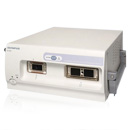
Miniature Probe
UM-DP20-25R

Miniature Probe

The UM-DP20-25R offers dual-plane reconstruction (DPR) scanning via the channel of a standard endoscope, making intraductal ultrasonography (IDUS) easier, and images simple to interpret. An automatic, 40 mm linear scanning stroke allows entire lesions to be scanned in one pass, making procedures more efficient.
When the miniprobe UM-DP20-25R is connected to the probe-driving unit MAJ-935, and ultrasound centres EU-ME2 or EU-M60 with 3D software, its DPR function allows acquisition of a helical ultrasound scan, which can be converted to a 3D ultrasound image. These images allow you to more easily define tumour volumes, invasion depth and the relationship between the lesion and surrounding vasculature. To make IDUS as practical and accessible as possible, this ultrasound miniature probe can pass through standard medical endoscope working channels as narrow as 2.8 mm.
Operating via a standard endoscope, the DPR function of this miniprobe allows helical ultrasound scanning, which can be converted into 3D images.
The transducer can be automatically pulled back 40 mm, creating a helical ultrasound scan. This allows entire lesions to be scanned in one pass.
This DPR miniature probe uses afrequency of 20 MHz, giving detailed, high-resolution ultrasound images of the region of interest.
When used in ‘standard’ mode, the DPR probe captures and displays radial and linear images simultaneously, allowing analysis in intricate detail.
This ultrasound miniature probe can be extended through endoscope working channels starting at just 2.8 mm, and its DPR function allows the generation of 3D ultrasound images, when used in conjunction with appropriate software.
The automatic scanning procedure retracts the transducer by 40 mm, capturing a helical ultrasound scan, while the miniprobe retains its position. This allows you to image whole lesions in a single pass, making procedures more efficient.
The scanning frequency of 20 MHz allows the acquisition of detailed, high-resolution ultrasound images during IDUS.
Radial or both radial and linear images can be displayed on a single monitor simultaneously with the probe in 'standard' mode, making it easier for you to interpret ultrasound images collected during IDUS.
| Display mode | B-mode |
|---|---|
| Scanning Direction | Perpendicular to the direction of insertion |
| Scanning Method | Mechanical radial/helical scanning |
| Scanning Field of View | 20 MHz |
| Contact Method | Direct contact method Sterile de-aerated water immersion method |
| Working Length | 2,050 mm |
| Total Length | 2,210 mm |
| Insertion Tube-Outer Diameter | 2.5 mm |

Contents end