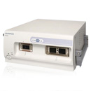
Radial Scanning Ultrasound Endoscope
GF-UE160-AL5

Radial Scanning Ultrasound Endoscope

The world's first 360° electronic radial scanning scope expands the potential of EUS, combining exceptional scope capability and maneuverability with advanced ultrasound functionality, enabling blood flow confirmation for easier orientation in the pancreatobiliary region.
Expanding the potential of EUS, this 360° electronic radial echoendoscope combines exceptional scope capability and maneuverability with superb ultrasound image quality and advanced ultrasound functionality.
The GF-UE160-AL5 enhances resolution and penetration depth, enabling confirmation of blood flow conditions for easier orientation in the pancreatobiliary region, and is compatible with the Olympus EU-ME2 ultrasound centre and Hitachi Aloka Alpha 5, 7, and 10 systems.
Providing a full 360° ultrasonic overview, enabling precise diagnostics of the GI tract and allowing an easier anatomical orientation via image rotation capability.
Five different frequencies, from 5 MHz up to 12 MHz, permit the highest image quality at every scanning area.
Built-in CCD enables crystal clear endoscopic video images of the targeted area with white light.
The small diameter, wide angulation range (130° up, 90° down/left/right) and short distal end of the echoendoscope enables excellent maneuverability in order to safely display all areas of interest.
Enables the investigation of vascular structures to characterise suspicious tissue findings and allow easier orientation within the pancreatobiliary region.
Clearly delineates lesion outlines and provides improved spatial and contrast resolution of the ultrasound image. THE further improves ultrasound image quality.
Allows the most precise tumour staging ever.
Five different frequencies (from 5 MHz up to 12 MHz), enables high resolution scanning.
Built-in CCD with a 100° field of view enables precise white-light diagnostics of the GI tract and easier orientation via image rotation capability.
The small diameter, wide angulation range (130° up, 90° down/left/right) and short distal end of the echoendoscope enables excellent maneuverability.
The lens cleaning function ensures the endoscopic field of view is clear at all times.
Usually used for EUS-guided FNA procedures, Doppler capability enables a detailed observation of blood flow within a lesion.
THE improves the ultrasound image quality without any need for contrast agents.
| Endoscopic functions | ||
| Optical System | Field of view | 100° |
|---|---|---|
| Direction of view | 55° forward oblique | |
| Depth of field | 3 to 100 mm | |
| Insertion Tube | Distal end outer diameter | Ø13.8 mm |
| Insertion tube outer diameter | Ø11.8 mm | |
| Working length | 1,250 mm | |
| Instrument channel | Channel inner diameter | Ø2.2 mm |
| Minimum visible distance | 3 mm | |
| Bending Section | Angulation range | UP 130°, DOWN 90°, RIGHT 90°, LEFT 90° |
| Total Length | 1,555 mm | |
| Lens Cleaning Function | Available | |
| Ultrasonic functions | ||
| Display mode | B-mode, M-mode, D-mode, Flow-mode, Powerflow-mode | |
| Scanning method | Electrical radial array | |
| Scanning direction | Perpendicular to insertion direction | |
| Frequency | 5/6/7.5/10MHz | |
| Tissue Harmonic Echo | 3.75S/3.75P/5.0R/5.0H | |
| Focusing Point | A maximum of 4 focusing points are available. (between F1 to F16) |
|
| Scanning range | 360° | |
| Contacting method | Ballon method Sterille de-aerated water immersion method |
|
| Image Rotation | Available | |

Contents end